
DETROIT – Wayne State University has added one of the best 3T magnetic resonance imaging (MRI) tools available to its arsenal of technologies to significantly enhance its research capabilities.
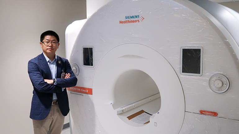
Wayne State is among the first institutions in the country to receive the new Siemens 3T MAGNETOM Cima.X scanner – a state-of-the-art, high resolution MRI system, designed to provide deeper insights into the human body.
Wayne State received a $2 million grant from the National Institutes of Health and an additional $1.38 million university match to purchase the scanner.
Equipped with the latest generation in imaging technology, the new scanner will be an asset for Wayne State researchers as they work to understand the progression of chronic diseases, evaluate the long-term effects of interventions and study the natural history of various conditions. The new technology can be a powerful tool for clinicians and scientists to detect threats early and prevent severe consequences, such as seeing coronary or vascular diseases before a heart attack or stroke or identifying prostate cancer in its earliest stage before a biopsy.
“The capabilities of the new MRI scanner will enable and support collaboration among faculty across the basic life and behavior sciences, translational clinical sciences and engineering,” said President Kimberly Andrews Espy, Ph.D. “The enhanced resolution and novel imaging capabilities advances transdisciplinary teaming that put us on the forefront of addressing complex medical challenges with cutting edge precision.”

Construction of a specialized laboratory space inside the Eugene Applebaum College of Pharmacy and Health Sciences was required for the new system, which was installed on May 17. It is expected to go live and hopefully scan its first human subject in mid-July.
The scanner will have an immediate impact on more than 18 research programs across Wayne State, ultimately enabling new discoveries into brain structure and function to help better understand the workings of the brain and etiology of disease. Wayne State researchers are studying neurological conditions and disorders like multiple sclerosis and Parkinson’s disease, as well as other important research areas such as the impact of stress, trauma and substance use on brain function. These and other research studies will greatly benefit from the investment in the new MRI system and its state-of-the-art features.

Wayne State has a long history of collaboration with Siemens in MR research, dating back to the early 2000s when Professor Mark Haacke, Ph.D., former director of the MR Research Facility, invented a technique called Susceptibility Weighted Imaging (SWI). Today, SWI is an essential brain MRI scan used in every Siemens MRI scanner, benefiting millions of individuals worldwide.
“We are thrilled to continue our partnership with Siemens as one of the first institutions in the country to receive this newly FDA-approved 3T MRI scanner,” said Yongsheng Chen, Ph.D., associate professor of neurology and director of the MR Research Facility. “This new model features an unparalleled gradient system that advances the boundaries of imaging, turning the invisible into the visible.
“Serving the entire Wayne State research community, this state-of-the-art scanner will enable us to remain at the forefront of neuroscience research and maintain our leadership in MRI research for the next 15 years and beyond.”
Research cores are a critical component to the research enterprise that offer state-of-the art equipment as a central hub of discovery.
“Research core facilities offer investigators cutting-edge equipment that they would not otherwise have access to,” said Ezemenari Obasi, Ph.D., vice president for research at Wayne State University. “These centralized facilities are hubs of discovery that facilitate collaboration between researchers from different fields, and aid in accelerating scientific breakthroughs at a much faster pace. Cores such as this MRI core facility are critical to growing research enterprises, and I am pleased that we are able to provide this innovative equipment to our scientists.”
Information reported in this press release was supported by the National Institutes of Health under award number S10OD028724.