A study conducted at the Wayne State University School of Medicine on tissue samples and cultured cells demonstrated that SARS-CoV-2 can efficiently infect human conjunctival epithelial cells in the eye, producing innate immune response.
“We were also able to demonstrate that the beta variant of concern replicates more and induces a much higher inflammatory response,” Ashok Kumar, Ph.D., said in an interview with Ocular Surgery News.
Epithelial cells make up protective layers throughout the body, including the eye.
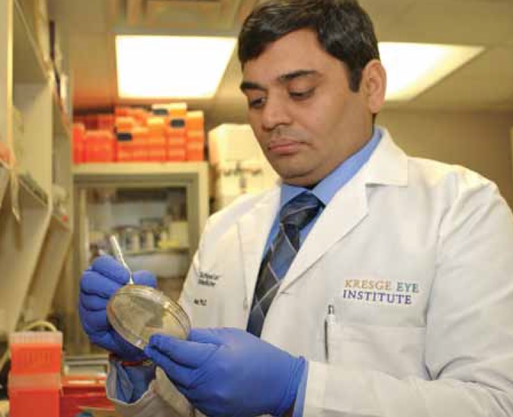
Dr. Kumar, an associate professor of Ophthalmology, Visual and Anatomical Sciences, appears on the cover of this month’s magazine, his second appearance since May 2021.
His research focuses on infectious diseases that affect the eye. In a previous article published in The Ocular Surface journal, Dr. Kumar and team identified the presence of SARS-CoV-2 RNA, and spike and envelope proteins, in postmortem corneal and scleral tissue of COVID-19 donors.
In the WSU team’s most recent study published in the same journal, “SARS-CoV-2 and its beta variant of concern infect human conjunctival epithelial cells and induce differential antiviral innate immune response,” similar findings were reported in postmortem conjunctival tissues. The mechanisms and timing of viral entry and replication, and the immune response to the virus, were clarified in a cultured human conjunctival epithelial cell, or HCEC, model.
The study was a collaboration between his lab at Wayne State, Cedars-Sinai Medical Center in Los Angeles and the University of California, Los Angeles.
“At Cedars-Sinai, Dr. Alexander Ljubimov, an expert in ocular surface diseases and cell cultures, harvested the conjunctival tissue from healthy donors and cultured the cells in a dish. At UCLA, Dr. Vaithi Arumugaswami performed the infection studies in a BSL-3 high-containment facility, where the cells were infected with a parental strain of SARS-CoV-2 – the original Wuhan virus – and with the beta variant of concern – the so-called South African variant. Samples were then sent to us, and we performed molecular biology analyses,” Dr. Kumar said.
The study demonstrated that the parental strain and beta variant were able to infect the hCECs, possibly by binding to the ACE2 and TMPRSS2 receptors, known as entry receptors for SARS-CoV-2. In addition, three other receptors — CD147, Axl and neuropilin-1 — implicated in viral entry, including SARS-CoV-2, were also found to be expressed in this experimental model.
“Distinct expression of all these receptors was detected in most cultured hCECs, as well as in the conjunctival epithelium ex vivo. The presence of permissive viral entry receptors in the conjunctiva potentially enables the virus to use the eye as a site of replication and dissemination to other organs, as seen with other RNA viruses,” Dr. Kumar said.
Once infected, the innate immune response was triggered, leading to production of cytokines, chemokines, interferons and interferon-stimulated genes.
The study also showed that that virus replication, the expression of receptor genes and the immune response have an early peak at 24 hours and then decline. The trend was consistent in the cells derived from three different donor samples.
“SARS-CoV-2 infects the conjunctiva but does not kill the cells. The conjunctiva as well as the cornea seem to be able to mount antiviral response to the virus, and that could be the reason why we don’t see a much higher increase beyond 24 hours. Very different is the behavior of the virus in the lungs, where cells are killed in a very high number. The lungs may be the real primary target of SARS-CoV-2,” Dr. Kumar said.
The emergence of new variant strains of SARS-CoV-2 — more transmissible, more aggressive and potentially resistant to vaccines — is a concern and may threaten the progress made so far thanks to mass vaccination, he added.
In an ongoing study, a large number of samples – mainly corneal samples – have been collected for screening from eye banks in seven states.
“So far, we have analyzed more than 600 samples, and those which were positive for SARS-CoV-2 are being analyzed by next-generation sequencing. This would indicate whether a specific variant is prevalent in the ocular tissue and to clarify the mechanisms underlying the increased inflammatory response to these variants of concern,” Dr. Kumar said.
Further research may find whether and to what extent SARS-CoV-2 can enter the system through the eye, and whether ocular involvement is secondary to systemic infection. Identifying organs and cell types permissive to viral entry and replication could help to develop preventive or therapeutic strategies against SARS-CoV-2 transmission, Dr. Kumar said.
The study was supported in part by National Institutes of Health grants R01EY026964 and R01EY027381 to Dr. Kumar, and an unrestricted grant to the Kresge Eye Institute and Department of Ophthalmology, Visual and Anatomical Sciences from Research to Prevent Blindness. The authors would like to thank Onkar Sawant, Ph.D., director of Research at Eversight, and organ procurement organization partners of Eversight, for contributing donated ocular tissues.