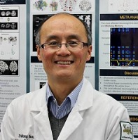
Zhifeng Kou, Ph.D., Wayne State University assistant professor of Biomedical Engineering and Radiology, found himself appalled at the limited methods neurosurgeons had to monitor traumatically injured brains. When a severely sick patient with evidence of a brain injury needed brain activity monitoring, a hole had to be drilled in the head and a long, thin catheter inserted into the brain — essentially sticking a pin into the brain and watching for electronic sign of abnormality. The process takes a relatively long time and sometimes must be repeated to locate the precise area of injury.
It seemed to Dr. Kou that a faster and less-invasive method of diagnosing traumatic brain injury could be developed using the magnetic resonance imaging he already worked with. His research concept received a $425,000 grant in October from the National Institutes of Health for a study that will investigate moderate to severe TBI effects in up to 30 intensive care patients at Detroit Receiving Hospital.
Teaming with Mark Haacke, Ph.D., director of the MR Research Facility and professor of Radiology, Dr. Kou is developing MRI-based techniques to show what is happening with veins deep inside the brain. Arteries deliver oxygenated blood, and veins drain the oxygen-depleted blood back to the heart and lungs. By getting a picture of what the veins are doing inside the skull, doctors can localize areas of the brain that are damaged and starved of oxygen. The veins themselves become a virtual brain catheter. By combining evidence of arterial blood supply with venous drain, Dr. Kou can detect brain regions with abnormal metabolic activities.
"Using the MRI, and without creating a hole, we can sense what part of the brain is hungry for blood, determine the real problem and give better support for the patient," Dr. Kou said. "These veins work exactly like a brain catheter in that they sense the surrounding tissue, except they're everywhere."
The perfusion and drainage pattern shown in an MRI can be an important tool for neurosurgeons, who cut the brain to effect a cure, and for the neurointensivist, who provides critical care for brain trauma patients. While Dr. Kou is not directly involved in treatment, he said the technique can give doctors valuable information on brain metabolism at different regions and guide them for proper management and treatment protocol. Should a brain catheter need to be placed for continuous monitoring, the MRI process can remove placement guesswork and provide an exact target location.
"The brain is very complex, and different structures within the brain have different physical attributes. Using this technique, we can develop a cerebral metabolic index that tells us which part of the brain is abnormal and which is normal," Dr. Kou said.
In addition to being noninvasive, the MRI delivers quick results. Traditional catheterization requires intervention by a brain surgeon and time both to install the catheter and wait for the area of the brain it's inserted into to stabilize and show normal activity. With the MRI, "you see the beautiful picture right away."
As an offshoot of Dr. Kou's project, his M.D.-Ph.D. student Natalie Wiseman will use the same technology to measure brain metabolic changes after a mild concussion. Wiseman received an F30 Ruth L. Kirschstein National Research Service Award for $224,000 toward her study, which is meant to identify which concussion victims are likely to experience slower recoveries. Wiseman is the first student from Dr. Kou's lab to receive such an award.
Dr. Kou's investigation will follow TBI patients from initial diagnosis through recovery, with additional analysis at the six-month mark. He hopes it will lead to a powerful new imaging tool to help doctors achieve the best possible patient recoveries.
"I'm a simple and straightforward person, and I just want to make something that physicians can understand and easily use," he said.
Dr. Kou's project includes Dr. Haacke as co-principal investigator as well as Gregory Norris, M.D., medical director of the Neurotrauma and Critical Care unit at Detroit Receiving Hospital; neuropsychologist John Woodard, Ph.D.; and radiologist Conor Zuk, D.O.