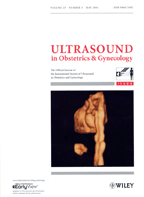
Wayne State University 's Dr. Jimmy Espinoza captured the cover of the May Journal of Ultrasound in Obstetrics and Gynecology with a novel three/four-dimensional ultrasound rendering of a fetal heart.
His featured article describes a novel rendering algorithm called the inversion mode with which he has obtained more accurate views of the venous connections of the fetal heart, allowing diagnosis of abnormalities. The inversion mode is a sonographic modality that transforms translucent heart and vascular structures into echogenic voxels, allowing them to become visible through ultrasound. Other fluid-filled structures including the stomach and bladder can also be seen as echogenic structures with this new rendering modality.
Using three- and four-dimensional ultrasound, volumes are obtained and analyzed to visualize veins that may range from only 1 to 4 mm in the fetus which are difficult to see with two-dimensional ultrasound. "This new rendering modality allows us to see more anatomical detail than before and could potentially improve the prenatal diagnosis of abnormal systemic venous connections to the fetal heart," Dr. Espinoza said.
Co-authors on the study include Drs. Luis Gonçalves, Wesley Lee, Moshe Mazor and Roberto Romero.